Derm Forge
Built at Meta Global AI Developer Hackathon · Oct 26, 2026 · Remote
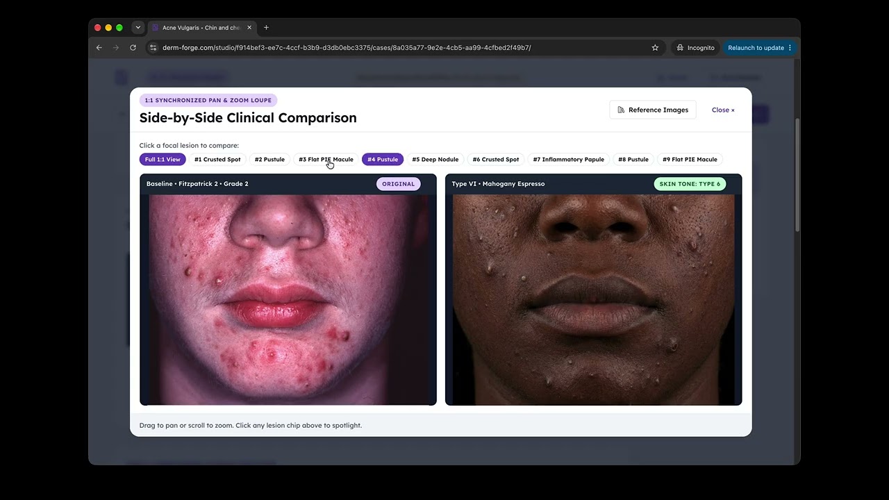
Derm Forge is a clinical case and optical simulation studio for medical faculty, built from scratch during the hackathon on Meta's API (muse-spark-1.3, sam-3.1, muse-image-1.0, and muse-wonder-1.0). Derm Forge is an educational authoring tool for faculty, not a patient-facing diagnostic or medical advice app. The physician sets the ground-truth diagnosis, baseline Fitzpatrick skin type, and severity grade upfront. THE PROBLEM Medical training images overwhelmingly depict fair skin (Fitzpatrick Types I and II). Because clinicians are undertrained on how melanin masks pink vascular redness in darker skin (Types IV to VI), patients face severe healthcare disparities: Black patients are over 3x more likely to receive a late-stage melanoma diagnosis (52% vs. 16%), driving a 70% vs. 94% 5-year survival gap, alongside everyday diagnostic delays in primary care. As CTO of Derm for Primary Care (where we train thousands of Nurse Practitioners annually), I have seen firsthand why static clinical photo libraries cannot fix this: 1. Educators never have the exact same patient and lesion geometry across all six Fitzpatrick skin tones to teach how the exact same lesion shifts optically across skin colors. 2. Educators never have controlled longitudinal follow-ups of the same patient showing +1 grade worsening, 6-week partial clearance, 16-week remission, flat post-inflammatory macules, and concave scars. THE SOLUTION I started with primary care's highest-volume condition and hardest optical stress-test: Acne Vulgaris (where up to 13 co-existing 3D and 2D lesion types present simultaneously). In skin of color, masked redness causes providers to either under-treat deep nodules until permanent scarring occurs, or mistake flat resolved hyperpigmentation (PIH) for active acne and needlessly prescribe oral antibiotics. Derm Forge transforms a single clinical photo into a complete NP training module across four chained stages: Stage 1 (PHI Redaction & Lesion Grounding): muse-spark-1.3 and sam-3.1 redact patient identifiers and snap pixel-tight boxes around focal lesions using grade- and skin-type-specific optical targets. Stage 2 (Cross-Skin-Tone Studio): muse-image-1.0 translates the baseline photo across Fitzpatrick Types I to VI while locking 100% of pore texture and 1:1 lesion geometry, paired with a 1:1 Synchronized Pan & Zoom Loupe and real-world clinical reference photos. Stage 3 (Disease Progression & Clearance): muse-image-1.0 maps Stage 1's boxes onto a 3x3 anatomical salience grid to simulate worsening, partial response, complete clearance, flat 2D PIE/PIH macules, and 3D concave scars. Stage 4 (Board Vignettes): muse-spark-1.3 and muse-wonder-1.0 author spoiler-free AANP/ANCC board flashcards bound to the generated images and numbered lesions, ready to deploy to the thousands of Nurse Practitioners we train each year.